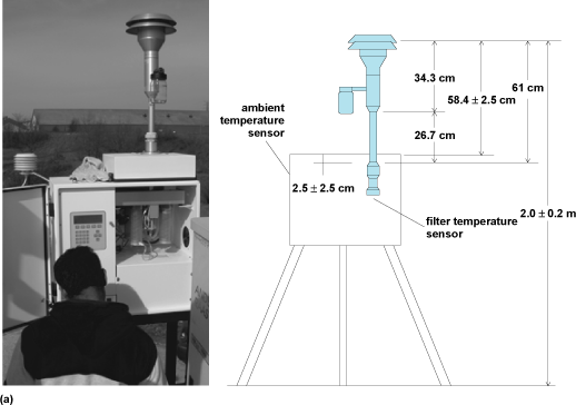
Environmental engineers are generally concerned with two types of air pollutants: gases and particulate matter (PM). Generally, the size categories of PM measured are (1) ≤2.5 μm in diameter and (2) 2.5–10 μm in diameter. Measurements are taken by instruments called dichotomous samplers, with components that use size exclusion mechanisms to segregate the mass of each size fraction (Fig. 1). Particles with diameters ≥10 μm are of less concern, since their mass is sufficiently large that they rarely travel long distances. Occasionally they are measured if a large particulate emitting source, such as a coal mine, is nearby.
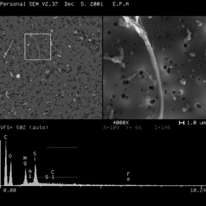
Particles that are highly elongated are usually differentiated as fibers. Such elongation is expressed as a particle's aspect ratio; that is, the ratio of the length to width. Environmentally important fibers include fiberglass, fabrics, and minerals (Fig. 2). Exposure to fiberglass and textile fibers is most common in industrial settings. Health problems have been associated with textile workers who were exposed to fibrous matter in high concentration for many years. For example, chronic exposure to cotton fibers has led to the ailment byssinosis, also known to as brown lung disease, which is characterized by narrowing of the lung's airways. When discussing fibers, it is likely that first contaminant to come to mind is asbestos, a group of highly fibrous, naturally occurring minerals with separable, long and thin fibers (Fig. 2).
Fibers are defined by convention, including by regulatory agencies such as the U.S. Environmental Protection Agency and Occupational Safety and Health Administration, to have aspect ratios equal to or greater than 3:1. Methods typically used to collect ambient air samples, such as the system shown in Fig. 1, are not suitable for collection of most fibers for subsequent analysis. Instead, asbestos fibers are typically collected using open-faced cassettes loaded with membrane filters. The Teflon® filter shown in Fig. 1 is not used for asbestos since no solvent is available to extract the fibers. Quartz filters cannot be used since they are made of mineral fibers, which are difficult to differentiate from asbestos fibers. The asbestos fibers are occluded by the larger-diameter quartz fibers as well. This is why mixed cellulose ester and polycarbonate filters in cassettes are used.
Asbestos fibers are strong and flexible enough to be spun and woven. They are resistant to heat and chemicals, making them useful for many industrial purposes. Because of their durability, asbestos fibers that get into lung tissue will remain there for long periods of time.
Fiber type and health effects
There are two general types of asbestos: amphibole and serpentine. Some studies show that amphibole fibers stay in the lungs longer than the serpentine mineral, chrysotile, and this tendency may account for their increased toxicity.
Generally, health regulations classify asbestos into six mineral types: chrysotile (the only asbestos member of serpentine group), which has long and flexible fibers, and five amphiboles, which have brittle crystalline fibers. The amphiboles include actinolite asbestos, tremolite asbestos, anthophyllite asbestos, crocidolite asbestos, and amosite asbestos. Each of the amphiboles includes the term asbestos because, to geologists, amphiboles can also have nonasbestiform habits (that is, the host rock is an amphibole that may contain a mixture of nonfibrous particles, fibers, and cleavage fragments).
The most important risk factors for asbestos-related diseases are length of exposure, air concentration of asbestos during the exposure, and smoking. Cigarette smoking and asbestos exposure are synergistic; that is, the risk of disease is multiplied if a person exposed to asbestos is a smoker (Table 1). There is an ongoing scientific debate about the differences in the extent of disease caused by different fiber types and sizes. Some differences may be due to the physical and chemical properties of the different fiber types. For example, several studies suggest that amphibole asbestos types (tremolite, amosite, and especially crocidolite) may be more harmful than chrysotile, particularly for mesothelioma. Other data indicate that fiber size (length and diameter) are important factors for cancer-causing potential. Some data indicate that fibers with lengths greater than 5.0 μm are more likely to cause injury than those less than 2.5 μm in length. Additional data indicate that short fibers can contribute to injury. This appears to be true for mesothelioma, lung cancer, and asbestosis. However, fibers thicker than 3.0 μm are of lesser concern, because they have little chance of reaching the lower regions of the lung.
Some groups of people who have been exposed to asbestos fibers in drinking water have higher-than-average death rates from cancer of the esophagus, stomach, and intestines. However, it is very difficult to tell whether this is caused by asbestos or other causes.
|
Exposure group |
Asbestos dose (fibers m−3) |
||||
|
8 |
50 |
80 |
500 |
2000 |
|
|
Male smokers |
1 (0–9) |
6 (0–55) |
9 (0–88) |
55 (0–550) |
221 (0–2210) |
|
Female smokers |
1 (0–5) |
2 (0–25) |
5 (0–41) |
25 (0–250) |
101 (0–1010) |
|
Male nonsmokers |
1 (0–1) |
1 (0–8) |
1 (0–11) |
8 (0–75) |
29 (0–290) |
|
Female nonsmokers |
1 (0–1) |
1 (0–3) |
1 (0–5) |
3 (0–28) |
11 (0–110) |
source: California Air Resources Board, Staff Report, Initial Statement of Reasons for Rulemaking, 1986.
Routes of exposure
Ambient air concentrations of asbestos fibers are about 10−5–10−4 fibers per milliliter (fibers mL−1), depending on the location. Prolonged human exposure to concentrations much higher than 10−4 fibers mL−1 is suspected of causing adverse health effects. Asbestos fibers are very persistent and resist chemical degradation (that is, they are inert under most environmental conditions) so their vapor pressures are nearly zero (they do not evaporate), and they do not dissolve in water. However, segments of fibers do enter the air and water when asbestos-containing rocks and minerals are weathered naturally or extracted during mining operations. One of the most serious exposures occurs when manufactured products, such as pipe wrapping and fire-resistant materials, begin to degrade or are improperly handled during renovation or removal activities. Small-diameter asbestos fibers may remain suspended in the air for a long time and be transported advectively by wind or water, before sedimentation. Like spherical particles, heavier asbestos fibers settle more quickly. Asbestos seldom moves substantially via soil. The fibers are generally not broken down to other compounds in the environment and will remain virtually unchanged over long periods.
Although most asbestos is highly persistent, chrysotile, the most commonly encountered form, may break down slowly in acidic environments. Asbestos fibers may break into shorter strands, and therefore into increased number of fibers by mechanical processes (such as grinding and pulverization). Inhaled fibers may get trapped in the lungs and with chronic exposures build up over time. Some fibers, especially chrysotile, can be removed from or degraded in the lung with time.
Because of its toxicity, it is necessary to have effective monitoring techniques and analytical methods to detect, quantify, and control asbestos in the environment. A number of techniques and methods have been developed to detect and quantify asbestos in bulk samples, air, water, settled dust, and soil.
Analytical methods
Numerous methods are used to measure asbestos: asbestos and mineral fibers in air by scanning electron microscopy (SEM), scanning transmission electron microscopy (STM), asbestos and mineral fibers in air by phase-contrast microscopy, asbestos and mineral fibers in bulk materials, manufactured mineral fibers, asbestos in soils, asbestos in dust, and asbestos in water.
The most widely used techniques are based primarily upon observing the fiber by either optical or electron microscopy. These techniques include phase-contrast microscopy (PCM), polarized light microscopy (PLM), scanning electron microscopy, and transmission electron microscopy. Some methods also use x-ray diffraction spectrometry (XRD) for identifying mineral phases and quantitative analysis. Qualitative and quantitative analysis of fibers by each technique are discussed in Table 2.
|
Analytical method |
Qualitative analysis |
Quantitative analysis |
|---|---|---|
|
Phase contrast microscopy (PCM) |
PCM is used for monitoring airborne levels of asbestos in workplace environments where the fiber type is known. PCM can detect fibrous materials but cannot directly distinguish asbestos fibers from other fibrous materials. Air samples are collected on a cellulose ester membrane filter with 0.45–1.2 μm pore size. A portion of the filter is mounted on a microscope slide, cleared using an organic solvent, and fibers are observed in a bright field at a magnification of 100 to 400×. PCM detects fibers as thin as 0.25 μm in thickness. |
PCM methods report fibers per cubic centimeter of air sampled (f cm−3). Normally fibers longer than 5 μm in length and having an aspect ratio (length-to-width) of 3:1 or greater are counted. Some protocols use an aspect ratio of 5:1 and count fibers with diameters less than 3 μm. It should be noted that current risk models are based on fibers with a 3:1 aspect ratio, and data from methods employing a 5:1 aspect ratio cannot be used by such models. |
|
Polarized light microscopy (PLM) |
PLM is used for determining asbestos and other fibers, and minerals in bulk samples. A portion of the bulk material is examined by stereomicroscopy at a magnification 10 to 60× and subsamples of the various phases in the material are taken and mounted on a microscope slide in various refractive index liquids. Fibers are examined at magnifications 100–500× and identified by their optical properties including morphology (characteristic shape), color, refractive indices, birefringence, extinction angle, and sign of elongation. Characteristic dispersion staining colors are commonly used to identify mineral type. |
PLM methods typically report fibers by percent area or percent weight. The percentage may be estimated by visual estimation, comparison with charts showing various area percentages or with gravimetrically prepared standards, or by a point counting procedure. Although point counting is a systematic means of determining the relative amounts of materials in a mixture, it is not a reliable means of determining mass since one point may represent a single thin fiber or a large mineral “boulder.” Results using these techniques are usually considered “semiquantitative” since they do not measure quantity directly. |
|
X-ray diffraction spectrometry (XRD) |
XRD is used to assist in identifying mineral phases including asbestos and may be used to estimate quantity of the mineral phase. An x-ray beam is directed to the sample and a diffraction pattern characteristic of the mineral phase is produced. The pattern is compared to standard patterns produced by known minerals by means of reference files or through computer analysis. XRD cannot distinguish between asbestiform and nonasbestiform materials of the same mineral phase. XRD results must be confirmed by PLM, SEM, or TEM. |
In addition to the qualitative identification, the intensity of the diffraction pattern indicates the amount of material present. A comparison of selected peak heights on the XRD diffractogram to standards of known amounts of a mineral phase may assist the analyst in quantitative analysis of a sample. |
|
Scanning electron microscopy (SEM) |
SEM may be used for analysis of fibers in air or in bulk materials (Figs. 2 and 4). Typically, air samples are collected on cellulose ester or polycarbonate membrane filters and prepared for analysis. An image of the surface features on the filter is generated and fibers may be observed and counted. SEM magnifications may range from 2000 to 20,000× and higher. Fibers may be detected that are much thinner than those detected by optical microscopy such as PCM or PLM. An energy dispersive spectrometer EDS) unit, used in conjunction with the SEM, may detect the chemical composition of the fibers. |
Airborne fibers measured by SEM are typically reported in fibers per cubic centimeter. Fibers in bulk materials measured by SEM are typically reported as a percentage of the total sample with the percentage determination being made by visual estimation, comparison with standards of known composition, or gravimetry. Counting protocols for fibers by SEM is similar to those by TEM. |
|
Transmission electron microscopy (TEM) |
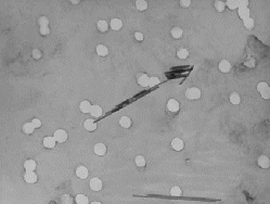
TEM may be used for analysis of fibers in air or in bulk materials (Fig. 3). Air samples collected on membrane filters or bulk samples transferred to membrane filters are carbon coated and a thin carbon layer including the fibers is transferred to a grid for analysis. TEMs provided magnifications from about 5000 to 20,000× or more and fibers with diameters of about 0.01 μm can be detected. An EDS unit can provide the chemical composition. In selected area electron diffraction (SAED), an electron beam is directed to the fiber, producing an electron diffraction pattern representing the crystal structure of the material. TEM is the most definitive technique for detecting and identifying asbestos materials. |
Over the years, a number of optical and electron microscopy methods have been developed to detect and quantify asbestos in air and other matrices. Each method has its strengths and weaknesses, and must be carefully evaluated to determine how best to detect and quantify asbestos under a given circumstance. Sampling efficiency is essential in exposure assessment.
Typically, mixed cellulose ester (0.45- or 0.8-μm pore size), and to a lesser extent capillary-pore polycarbonate (0.4-μm pore size) membrane filters are used to collect airborne asbestos for count measurement and fiber size analysis. The pore size specification for a membrane filter is an absolute specification only for capillary-pore-type filters, such as polycarbonate (PC). The pore size rating for tortuous-path filters (many entrapments), such as mixed cellulose ester (MCE) filters, is an effective pore size and not a specification that particles exceeding that size be retained by the filter.
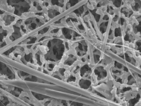
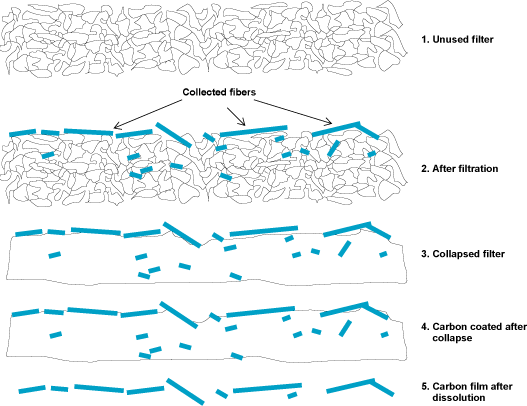
The two types of filters differ in their chemical and physical composition. Polycarbonate filters have a smooth filtering surface; the pores are cylindrical, almost uniform in diameter, and essentially perpendicular to the surface (Fig. 3). Because of their smooth surfaces, capillary-pore membrane filters are particularly useful for collecting particles that will be observed with a scanning electron microscope. A mixed-cellulose ester filter is a thicker filter with a spongelike appearance and relies on a tangled maze of cellulose ester strands to trap fibers (Fig. 4). For microscopic analysis of asbestos deposited on the filter, it is critical that the fibers be in a single plane to ensure they are in focus during analysis. This requirement is simple to achieve for PC filters because of the smooth filtering surface, whereas the MCE filter requires two additional steps in the direct preparation procedure (Fig. 5). The MCE filter must be collapsed with an organic solvent, and then the top layer of the collapsed filter material must be etched away with a low-temperature plasma asher.
Comparison of air filter types
Collection efficiency of the 0.45-μm pore size MCE filter for aerosols of asbestos fibers ≥0.5 μm in length is greater than that for a 0.8-μm pore size MCE filter (Table 3). This difference in collection efficiency is not maintained for asbestos structures longer than 5 μm. If the exposure study is focused on fibers <5.0 μm in length, the investigator should use MCE filters with a 0.45-μm pore size. If the exposure study exclusively focuses on structures longer than 5.0 μm, then either filter pore size may be used.
|
Mean concentration (structures/mm2) by filter |
||||
|---|---|---|---|---|
|
pore size and nominal loading |
||||
|
0.45 μm |
0.8 μm |
|||
|
Batch |
Low |
High |
Low |
High |
|
Fibers ≥0.5 μm |
||||
|
1 |
321 |
— |
274 |
— |
|
2 |
— |
958 |
— |
743 |
|
3 |
413 |
— |
388 |
— |
|
4 |
— |
1512 |
— |
1304 |
|
Fibers >5 μm |
||||
|
1 |
84 |
— |
80 |
— |
|
2 |
— |
373 |
— |
313 |
|
3 |
92 |
— |
100 |
— |
|
4 |
— |
301 |
— |
333 |
source: Data from the U.S. Environmental Protection Agency study of asbestos collection efficiency of mixed cellulose ester filters, 2007 (J. Kominsky, D. Vallero, and M. Beard, U.S. EPA Report, Comparison of Chrysotile Asbestos Collection Efficiencies on Mixed-Cellulose Ester Filters, 2007).
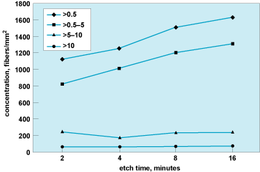
There is a significant difference in the effect of etching times for fibers <5.0 μm and those >5.0 μm in length (Fig. 6). The mean concentration of asbestos fibers ≥0.5 μm in length increases with etching time (2, 4, 8, and 16 min) of 0.45-μm pore size MCE filters. For fibers >5.0 μm in length, there is no significant difference in numbers of structures counted at the etching times used in these tests. Since most asbestos exposure risk models include fibers >5.0 μm in length, the 8-min etching time specified in ISO 10312:1995 is adequate. However, if an exposure study is focused on fibers <5.0 μm in length, the etching time of 8 min should be reviewed. Further study is needed to determine the etching time beyond which no significant increase in asbestos concentration of fibers <5.0 μm in length is expected.
[Disclaimer: The U.S. Environmental Protection Agency through the Office of Research and Development funded and managed some of the research described here. The present article has been subjected to the Agency's administrative review and has been approved for publication.]
See also: Amphibole; Asbestos; Chrysotile; Electron microscope; Environmental engineering; Mutagens and carcinogens; Phase-contrast microscope; Polarized light microscope; Respiratory system disorders; Scanning electron microscope; Serpentinite; X-ray diffraction


