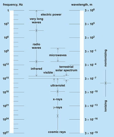
The study of the interactions of electromagnetic energy (usually referring to frequencies below those of visible light; Fig. 1) with biological systems. This includes both experimental and theoretical approaches to describing and explaining biological effects. Diagnostic and therapeutic uses of electromagnetic fields are also included in bioelectromagnetics.

Background
The interaction of electromagnetic fields with living organisms has intrigued both physicians and engineers since 1892 when J. A. d'Arsonval, a French physician and physicist, applied an electromagnetic field to himself and found that it produced warmth without muscle contraction. Subsequently, the use of electromagnetic energy to heat tissue became a common therapy, and in 1908 Nagelschmidt introduced the term diathermy to describe this process. During the 1930s “short-wave” diathermy (27 MHz) was in common use by physicians. World War II spurred the development of high-power microwave sources for use in radar systems. Shortly thereafter, concern over the safety of radar was voiced, leading to investigation of the biological effects of microwave radiation. Detailed study of the therapeutic potential of diathermy at microwave frequencies began after World War II as high-power equipment became available for medical and other civil applications.
Rapid growth in electronic systems for industrial, military, public service, and consumer use occurred during the 1970s and 1980s. Much of this equipment has the capability of emitting significant levels of electromagnetic radiation. The most extensive exposure to radio-frequency energy is from the thousands of transmitters authorized by the Federal Communications Commission (including commercial broadcast stations, cellular telephone transmitters, walkie-talkies, and microwave and satellite links). In addition, there are millions of unregulated citizen-band stations in use. The National Institute for Occupational Safety and Health estimates that more than 20,000,000 Americans are occupationally exposed to radio-frequency sources mainly from the heating and drying of plastics, textiles, wood products, and other manufactured goods. The use of electromagnetic fields in medicine increased as radio-frequency-induced hyperthermia was applied to cancer therapy. See also: Amateur radio
Energy absorption
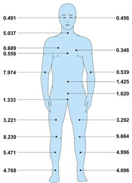
Electromagnetic energy is not absorbed uniformly across the geometric cross section of a biological organism. The total quantity of energy absorbed and the sites of maximum energy absorption depend on the frequency and polarization of the electromagnetic field, as well as on the electrical characteristics (the dielectric constant and conductivity—two properties of the tissue that control its interaction with electromagnetic radiation—which are frequency-dependent and vary with type of tissue), mass, and geometry of the absorbing object. In principle, the distribution of absorbed energy in an animal can be calculated from classical electromagnetic theory. However, the problem of energy distribution has not been solved for an object as complex as an animal. Simple calculations that assume the exposed system is of regular shape (for example, spheroidal) and of homogeneous composition allow some generalizations to be made. For an average person, maximal absorption (resonance) is predicted at approximately 70 MHz. When a complex target, such as a human being, is considered, resonant frequencies are also found in anatomically distinct portions of the body, such as head or leg. The internal energy absorption pattern has been calculated for several simple models: semi-infinite slab, homogeneous sphere, multishell sphere, and multiblock model of a human. While none of these models actually simulates a real human being, local regions of higher-than-average levels of energy absorption have been calculated (Fig. 2). Development of instruments that do not interfere with the field permit measurement of partial body resonances and the pattern of energy deposition within some experimental systems. See also: Absorption of electromagnetic radiation

Biological effects
The induction of cataracts has been associated with exposure to intense microwave fields. Although heating the lens of the eye with electromagnetic energy can cause cataracts, the threshold for cataract production is so high that, for many species, if the whole animal were exposed to the cataractogenic level of radiation, it would die before cataracts were produced.
In 1961, it was reported that people can “hear” pulsed microwaves at very low averaged power densities (50 microwatts/cm2). It is now generally accepted that the perceived sound is caused by elastic-stress waves which are created by rapid thermal expansion of the tissue that is absorbing microwaves. The temperature elevation occurs in 10 microseconds, so that the rate of heating is about 1.8°F/s (1°C/s). However, the temperature increase is only about 0.00009°F (0.00005°C).
There are reports that microwave irradiation at very low intensities can affect behavior, the central nervous system, and the immune system, but many of these reports are controversial. Animals exposed to more intense electromagnetic fields that produce increases in body temperature of 1.8°F (1°C) or higher (thermal load equal to one to two times the animal's basal metabolic rate) demonstrate modification of trained behaviors and exhibit changes in neuroendocrine levels. Exposure of small animals to weaker fields has been shown to produce some changes in the functioning of the central nervous system and the immune system. While the mechanism is not yet known, a thermal hypothesis is not ruled out. Reports of the effect of very low-level, sinusoidally modulated electromagnetic fields on excitable cell and tissue systems have raised questions about the basic understanding of how those systems function.
Exposure of pregnant rodents to intense electromagnetic fields can result in smaller offspring, specific anatomic abnormalities, and an increase in fetal resorption. However, fields that produce significant heating of the gravid animal have been shown to be teratogenic.
Most biological effects of microwaves can be explained by the response of the animal to the conversion of electromagnetic energy into thermal energy within the animal. However, a few experiments yield results that are not readily explained by changes of temperature.
In an industrial society, an appreciation of the effects of stationary electric and magnetic fields, and of extremely low-frequency fields are important because of the ubiquitous nature of electricity. When the body is in contact with two conductors at different potentials, current flows through it. Typical adult-human thresholds for 50- or 60-Hz currents are:
Reaction Total body current
Sensation 1 mA
“Let go” 10 mA
Fibrillation 100 mA
The “no contact” case, such as that experienced by an individual or animal under a high-tension transmission line (the field strength directly under a 765-kV line is 4–12 kV/m), has been investigated under controlled experimental conditions.
The possibility of hazard from occupational exposure to 50- or 60-Hz electric and magnetic fields has not been documented and is a subject of debate. Epidemiological studies have been undertaken to determine the health implications for workers and the general public exposed to these possible hazards. In addition, laboratory experiments have been designed to study the interactions of electric and magnetic fields with biological systems.
Some bacteria swim northward in stationary magnetic fields as weak as 0.1 gauss (the Earth's magnetic field is about 0.5 gauss at its surface). These bacteria contain iron organized into crystals of magnetite. Magnetite is also found in the brains and Harderian glands of birds that can use local variations in the Earth's magnetic field for navigation and orientation. See also: Biomagnetism; Microwave; Radiation biology
Mechanisms
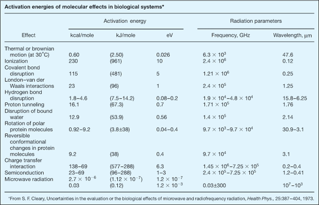
The energy associated with a photon of radio-frequency radiation is orders of magnitude below the ionization potential of biological molecules (see table). Covalent bond disruption, London–van der Waals interactions, and hydrogen-bond disruption, as well as disruption of bound water or reversible conformational changes in macromolecules, all require more energy that is contained in a single microwave photon. There is a possibility that absorption of microwaves (or slightly shorter millimeter waves) may produce vibrational or torsional effects in biomacromolecules. Thus, if radio-frequency electromagnetic fields have a specific action on molecules that alter their biological function, it will be through a route more complicated and less understood than that associated with ionizing radiation.

The most common mechanism by which electromagnetic fields interact with biological systems is by inducing motion in polar molecules. Water and other polar molecules experience a torque when an electric field is applied. In minimizing potential energy, the dipole attempts to align with the ever-changing electric field direction, resulting in oscillation. Both free and oriented (bound) water undergo dielectric relaxation in the radio-frequency region. The excitation of water, or other polar molecules, in the form of increased rotational energy is manifest as increased kinetic energy (elevation of temperature), but molecular structure is essentially unaltered if elevations are not excessive.
Alternating electromagnetic fields can cause an ordering of suspended particles or microorganisms. This effect, often called pearl-chain formation because the particles line up like a string of beads, results from a dipole-dipole interaction. Nonspherical particles may also be caused to orient either parallel or perpendicular to an applied field. These effects have been observed only at a very high field strength.
Electromagnetic energy absorbed by biological material can be converted into stress by thermal expansion. This phenomenon is caused by a rapid rise of temperature either deep within or at the surface of the material, and thus creates a time-varying thermal expansion that generates elastic stress waves in the tissue.
Medical applications
The therapeutic heating of tissue, diathermy, has been used by physicians for many years. “Short-wave” diathermy has been assigned the frequencies 13.56, 27.12, and 40.68 MHz, while microwave diathermy has been assigned 915, 2450, 5850, and 18,000 MHz. Short-wave diathermy provides deeper, more uniform heating than does diathermy at higher frequencies. See also: Thermotherapy
High-intensity radio-frequency fields have been used to produce hyperthermia in cancer patients. If a part of the body containing a tumor is heated, the difference of temperature between tumor and surrounding tissue can be increased, at times producing elevations to tumoricidal temperatures of 110–122°F (43–45°C) within the tumor while the surrounding tissue is below the critical temperature at which normal cells are killed. In addition, radio-frequency hyperthermia is often used in combination with x-ray therapy or with chemotherapy. In these cases, the tumor temperature is kept at 114–116°F (41–42°C) to enhance the effectiveness of radiation or chemotherapy.
The development of bone tissue (osteogenesis) can be stimulated electrically either with implanted electrodes or by inductive coupling through the skin. The noninvasive technique uses pulsed magnetic fields to induce voltage gradients in the bone. This therapy has been used successfully to join fractures that have not healed by other means.
Electromagnetic fields were first used for medical diagnosis in 1926 when the electrical resistance across the chest cavity was used to diagnose pulmonary edema (water in the lungs). At frequencies below 100 kHz, the movement of ions through extracellular spaces provides the major contribution to conductivity through the body. Thus, fluid-filled lungs can be detected by their lower resistivity. See also: Radiology
Another diagnostic tool, magnetic resonance imaging, uses the behavior of protons, or other nuclei, in an electromagnetic field to obtain imagesof organs in the body. These images are as good as, and sometimes better than, those obtained with x-ray computed tomography. Magnetic resonance imaging is especially useful when viewing the head, breast, pelvic region, or cardiovascular system. In magnetic resonance imaging, a static magnetic field is applied to align nuclear magnetic moments, which then precess about the field direction with a characteristic frequency. When a radio-frequency field is applied transverse to the direction of the magnetic field, nuclei are driven to the antiparallel state. As they return to the ground state, they radiate at their resonant frequency. The strength of the signal is proportional to the concentration density of the nuclei being studied. Other information for image analysis can be obtained from the relaxation time constants of the excited nucleus. See also: Computerized tomography; Medical imaging
Internally generated fields associated with nerve activity (electroencephalography) and with muscle activity (electrocardiography, magnetocardiography) are used to monitor normal body functions. There may be other uses of electric currents or fields in growth differentiation or development which have not yet been explored. See also: Cardiac electrophysiology; Electroencephalography; Electromagnetic radiation; Electromyography
Electric fields now play a role in biotechnology. Intense, pulsed electric fields (about 60,000 V/m) produce short-lived pores in cell membranes by causing a reversible rearrangement of the protein and lipid components. This permits the entrance of deoxyribonucleic acid (DNA) fragments or other large molecules into the cell (electroporation). The fusion of cells using electric fields (electrofusion) is easy to control and provides high yields. A weak, nonuniform alternating-current field is used to bring the cells into contact (dielectrophoresis), and then a brief, intense direct-current field is applied, causing the cell membranes to fuse.


